

In colposcopy, there will be times when we face the need to perform cone biopsies or a difficult loop electrosurgical excision procedure (LEEP). Navigating cone biopsy and difficult LEEP cases requires a combination of meticulous planning and refined techniques.1
Identifying the “Difficult LEEP”
A number of clinical factors can make a LEEP more technically challenging. A large transformation zone or wide ectropion may make it difficult to remove the full area of abnormal epithelium in a single pass. Vaginal wall laxity can further compromise exposure by obscuring the cervix, while postmenopausal vaginal narrowing and atrophy may limit access, reduce visualisation, and increase the risk of trauma or bleeding. Previous excisional treatment may distort cervical anatomy through scarring. When the anterior or posterior fornix is obliterated, the cervix may sit flush with the vagina, leaving less space between the cervix and adjacent structures. This increases the technical difficulty of excision and raises the risk of inadvertent injury to nearby tissue.
Patient-specific factors are also important. In very young patients, particularly those wishing to preserve fertility, there is a need to balance adequate excision for treatment with preservation of as much normal cervical tissue as possible. Anxiety and discomfort may add to the difficulty of the procedure.
The Three Ps of Setting Up for a Difficult LEEP
Success when performing a difficult LEEP begins long before the LEEP wire touches the tissue. A robust set-up is non-negotiable and should utilise the three Ps: patient, position, and preparation.
1. Patient
- Consider if the procedure is best performed under local anaesthetic or general anaesthetic (GA). For example, if the patient is anxious, GA may be more suitable.
- The patient should be adequately counselled and have expectations managed, ensuring that they are well prepared for the procedure.
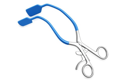
Fig. 1. Lateral vaginal wall retractor.
2. Position
- Positioning and visualisation of the cervix is critical for performing a safe and successful LEEP.
- Use of lateral wall retractors (Figure 1) may be helpful to optimise exposure.
- Surgeons should take advantage of the dynamic nature of the speculum and adjust its position throughout the procedure to maintain the best possible view and access to the cervix.
3. Preparation
- Involve an experienced surgeon when complexity is anticipated.
- Perform colposcopy to identify the TZ and lesions with acetic acid and Lugol’s iodine. Plan your LEEP procedure.
- Be diligent in checking your equipment set-up, ensure you have a functioning smoke extractor, and choose an appropriate size LEEP loop. This will depend on the size of your TZ and your lesion.
- Ensure availability of local anaesthetic. We prefer Lignospan (2% lidocaine injection with epinephrine).
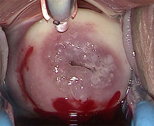
Fig. 2. Local anaesthetic infiltration with
blanching effect.
Refining the LEEP Technique
1. Local anaesthetic
Using two ampules of Lignospan, a practical approach is to infiltrate ½ ampule at the three and nine o’clock positions on the cervix, just laterally to the TZ (blanching the surface first, then advancing deeper), followed by ½ ampule each at 12 and six o’clock positions on the cervix, again lateral to the TZ (Figure 2).
The blanching technique involves introducing the needle 1-2mm into the cervical stroma. This does more than provide anaesthesia. The added volume expands the surrounding normal cervical tissue, in effect creating more working space by displacing the TZ medially, which can make the target area easier to access with the LEEP loop for excision while moving it away from the surrounding vaginal wall, avoiding risk of inadvertent injury.
Always warn the patient that they may experience tachycardia or lower limb muscle trembling due to the epinephrine component in the local anaesthetic.
2. Check your diathermy settings
Ensure use of the yellow button (cut) or blend rather than the blue (coagulation) during a LEEP procedure.2 Avoid dragging the LEEP wire through cervical tissue, as this can cause excessive charring and increase the risk of the loop becoming caught in the cervical stroma mid-procedure. Instead, apply current before tissue contact and glide the loop through the tissue in one smooth motion, using the cutting spark to achieve a more precise excision.2
Diathermy settings should be tailored to the loop size because it changes how electrical current is delivered.3 In practice, settings of 50/50 may be used for a 20 × 8mm loop. Smaller loops, such as 10 × 8mm, settings of 30/30 may be more appropriate. Larger loops have a greater metal surface area, so the current is distributed more broadly, and higher power is usually needed to maintain an effective cutting effect. Smaller loops concentrate the current over a smaller surface area, so lower power settings help minimise unnecessary thermal artefact.
3. The excision
Aim for a U-shaped rather than C-shaped excision. A U-shaped excision avoids transection of the TZ and ensures the TZ is removed symmetrically (Figure 3). For large lesions, a three-pass technique is recommended: one large central pass followed by thinner peripheral passes (Figure 4).
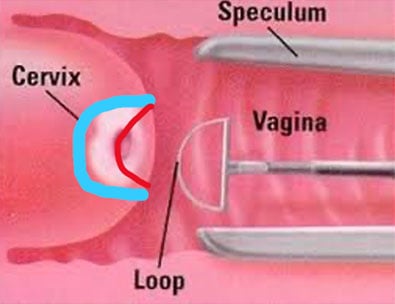
Fig. 3. Aim U-shaped excision (light blue) rather than shallow C-shaped excision (red).
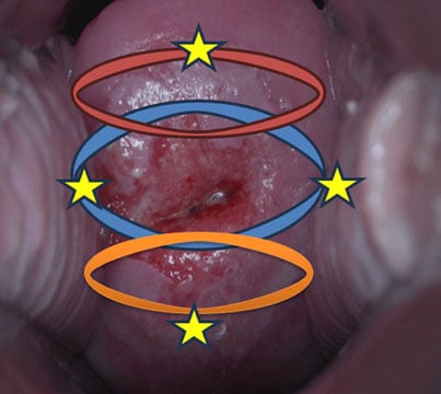
Fig. 4. For a large lesion, infiltrate edges of the TZ liberally with local anaesthetic to condense the TZ and create more working space. Use the three-pass technique. The first pass is the large central pass to adequately remove the majority of dysplasia (blue). The thinner anterior and posterior pass using only top half (red) and bottom half (orange) of LEEP loop to excise the rest of the lesion.
4. Preventing stenosis
Following a LEEP procedure, apply diathermy only to edges of the cervical defect to reduce the risk of post-operative cervical stenosis.
5. Managing excision depth carefully
During a LEEP, the cervix is often placed under significant tension with the speculum, which can make it appear flatter than it truly is. As a result, the depth of excision can easily be greater than intended. For this reason, it is often safer to aim for a shallower excision rather than risk going too deep. This is especially important in younger patients, where preserving as much normal cervical tissue as possible is a key part of reducing future obstetric risk.
Role of a Cone Biopsy
While LEEP is common, a cold-knife cone biopsy is indicated for specific high-risk scenarios where both diagnosis and treatment requires an intact specimen for histological evaluation, such as adenocarcinoma in situ (ACIS), microinvasive or early stage cancer, or in some cases when the upper extent of a lesion cannot be visualised (Type 3 TZ).4
A cone biopsy is usually performed under GA, with a scalpel, to remove the entire transformation zone and any suspicious cervical lesion for a detailed histological assessment.4 Its key advantage is that it provides a specimen with minimal diathermy artefact, allowing more accurate evaluation of the extent of the lesion and margin status.5 This is especially important when malignancy is suspected, as the accurate assessment of specimen margins is critical to guide subsequent management.
Should a Cone Biopsy be Called a Brick Biopsy?
Traditionally, a cone biopsy refers to the excision of the cervical TZ in a cone-shaped resection.4 In our practice, we find that this can transect the transformation zone prematurely. For this reason, our preference is to fashion the specimen in a more “brick-shaped” configuration, allowing a more complete excision of the TZ, while preserving the specimen integrity for pathological assessment (Figure 5).
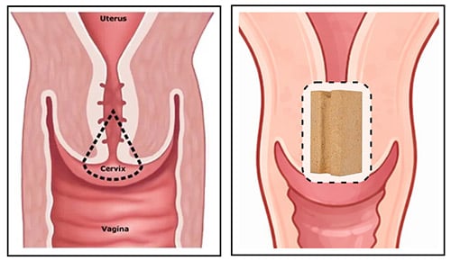
Fig. 5. Cone-shaped biopsy (left) may cause premature transection of TZ, while the brick-shaped biopsy (right) allows a more complete assessment and excision of the TZ, particularly a type 3 TZ.
The procedure sequence for a “brick biopsy” is as follows (Figure 6):
- Perform colposcopy and apply liberal amounts of local anaesthetic under non-sterile conditions.
- Under sterile conditions:
- Place Monaghan outside the TZ anteriorly (“no touch” technique to avoid disrupting the specimen).
- Place lateralised sutures at three and nine o’clock to ligate the cervical branch of the uterine arteries bilaterally and reduce bleeding.
- Apply iodine to assess extent of lesion on ectocervix.
- With a scalpel, incise the cervix to the appropriate depth, thereby fashioning the “walls” of your brick.
- Place sutures at the 12 and six o’clock positions on your specimen to provide traction.
- Excision is completed by fashioning the flat deep surface of the “brick biopsy”.
- Incorporate an endocervical curettage (ECC).
- Haemostasis with monsells and diathermy to edges. Our preference is to avoid Sturmdorf sutures as it increases the risk of cervical stenosis and unsatisfactory colposcopy at follow up.6
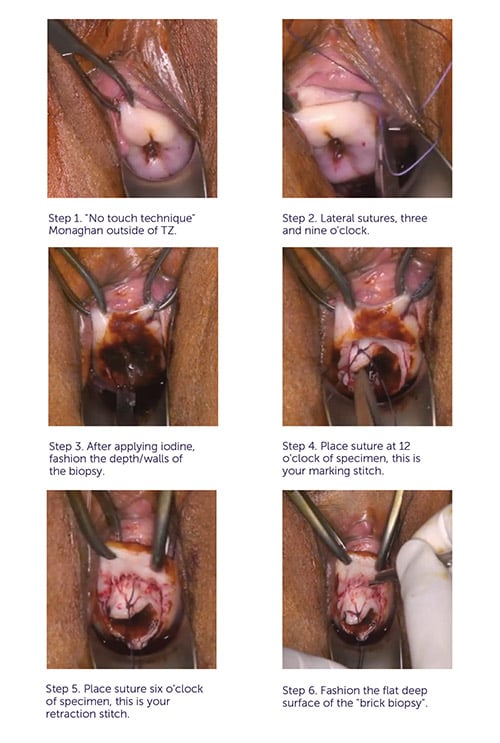
Fig. 6. Cone/Brick biopsy procedural image sequence.
Conclusion
Navigating the challenges of a difficult LEEP and cone biopsy requires planning. Whether the case demands a three-pass LEEP to manage a large lesion or a precision excision with a cone/brick biopsy for high-risk indications, success in these procedures stems from refined techniques and proactive prevention of complications.
The procedural approaches described here are adapted from teaching material developed by Dr Boo and A/Prof Pather for the Australian Society for Colposcopy and Cervical Pathology course.
References
- Boo, M., & Pather, S. Difficult LEEP and cone biopsy [PowerPoint slides]. Presented at the Australian Society for Colposcopy and Cervical Pathology Course, Sydney, NSW, Australia.
- Mayeaux, E. J., Jr, & Harper, M. B. Loop electrosurgical excisional procedure. The Journal of Family Practice.1993; 36(2): 214–219.
- Epperson, W. J., & Pfenninger, J. L. Electrosurgery for cervical intraepithelial neoplasia. Contemporary OB/GYN. contemporaryobgyn.net/view/electrosurgery-cervical-intraepithelial-neoplasia
- Cooper DB, Carugno J, Dunton CJ, et al. Cold Knife Conization of the Cervix. Updated 26 October 2023. StatPearls [Internet]. Treasure Island (FL): StatPearls Publishing. 2026. ncbi.nlm.nih.gov/books/NBK441845/)
- Krebs, H. B., Pastore, L., & Helmkamp, B. F. Loop electrosurgical excision procedures for cervical dysplasia: experience in a community hospital. American Journal of Obstetrics and G1993; 169(2 Pt 1):289–295. doi.org/10.1016/0002-9378(93)90078-w
- Gilbert, L., Saunders, N. J., Stringer, R., & Sharp, F. Hemostasis and cold knife cone biopsy: a prospective randomized trial comparing a suture versus non-suture technique. Obstetrics and G1989; 74(4):640–643.