

Background
Placenta accreta spectrum (PAS) is a rare pathology in pregnancy characterised by placental villi invasion onto, into, or through the uterine myometrium.1 The incidence of PAS in Australia and Aotearoa New Zealand is one in 2000 births and rates are increasing globally.1 Risk factors for PAS are previous caesarean birth, placenta previa, older maternal age, and multiparity.1 The optimal surgical management of PAS requires further research but may involve nontraditional uterine incision at caesarean hysterectomy to avoid the placenta.1,2 This report details a case of placenta accreta spectrum complicated by a placenta covering the entire anterior uterine surface.
Case Presentation
A 34-year-old woman with one previous caesarean birth presented with a preference for repeat caesarean birth for this pregnancy. Fetal morphology ultrasound at 20 weeks gestation revealed an anterior placenta praevia completely covering the internal os and overlying the caesarean scar with a posterior succenturiate lobe. An interval scan at 21 weeks showed a placenta overlying a thinned anterior myometrium without an apparent retroplacental clear space, raising concern for placenta accreta spectrum.
The patient presented acutely to our tertiary maternity unit at 23+1 weeks gestation with a painless antepartum haemorrhage provoked by intercourse. The haemorrhage was initially brisk but settling quickly, only requiring one menstrual pad overnight. Speculum examination revealed a closed cervix with slow fresh bleeding from the cervical os. Ultrasound now showed a 718g fetus with both estimated fetal weight and abdominal circumference above the 95th centile. There were several signs suggestive of placenta accreta spectrum. The placenta had become diffusely heterogenous with multiple vascular lacunae. The inferior placental margin was lobulated overlying a caesarean scar with thin myometrium measuring just 1mm. The posterior bladder wall was irregular, and large cervical vessels were seen potentially in continuity with the invasive placenta. Her bleeding remained settled, and she was discharged the next day with a plan to follow up in an outpatient antenatal clinic.

Fig. 1. Midline sagittal transvaginal ultrasound demonstrates a heterogenous placenta with vascular lacunae overlying a thin anterior myometrium measuring 1mm. Credit Radiology, Te Whatu Ora – Waitaha Canterbury.

Fig. 2. Transverse view on transvaginal ultrasound with colour Doppler demonstrates large cervical vessels at the irregular posterior edge of the bladder. Credit Radiology, Te Whatu Ora – Waitaha Canterbury.
Treatment
The patient re-presented at 23+6 after passing a 100mL clot without pain or provoking factors and was given a dose of intramuscular betamethasone. Bleeding continued in hospital up to 1L in total, and a magnesium sulphate infusion was started. She was given a dose of IV tranexamic acid and a red blood cell transfusion. Very preterm delivery became indicated when her ongoing haemorrhage reached two litres.
A bedside ultrasound scan was used for surgical planning, which revealed a cephalic baby with normal liquor and heart rate. There was anterior placenta praevia with anterior extension to the uterine fundus without visible window for uterotomy and fetal delivery, which had not been reported on prior imaging. Preoperatively, the patient was counselled about possible outcomes including leaving the placenta in situ with hysterectomy, leaving the placenta in situ without hysterectomy, or a very small chance of the placenta separating and leaving the uterus in situ.
Emergency caesarean hysterectomy was performed under general anaesthetic. The abdomen was entered by infraumbilical midline laparotomy. The lower uterine segment was ballooned. Large surface vessels covered the lower two thirds of the anterior uterine serosa. The decision was made for a midline vertical fundal incision in the uterus, which was extended toward the posterior aspect of the uterus. The fetus was delivered by breech extraction in an uncomplicated manner.
The umbilical cord was immediately clamped and cut. The maternal end of the cord was clamped close to the placenta and trimmed, then placed inside the uterus without attempting to deliver the placenta. The uterus was closed with a locking Vicryl suture, and bleeding at this site remained settled for the duration of the surgical procedure. Subtotal hysterectomy was performed, as this was the quickest procedure to remove the uterus and minimise blood loss. Surgicel haemostatic powder was placed at the cervical stump. Total estimated blood loss, including antepartum haemorrhage, was three litres. Four units of red cells and three units of cryoprecipitate were transfused in theatre. The neonate was transferred to the neonatal intensive care unit in expected condition.

Fig. 3. Anterior uterus after delivery of the fetus demonstrating large surface vessels. Credit Dr Olivia Smart, Te Whatu Ora – Waitaha Canterbury.
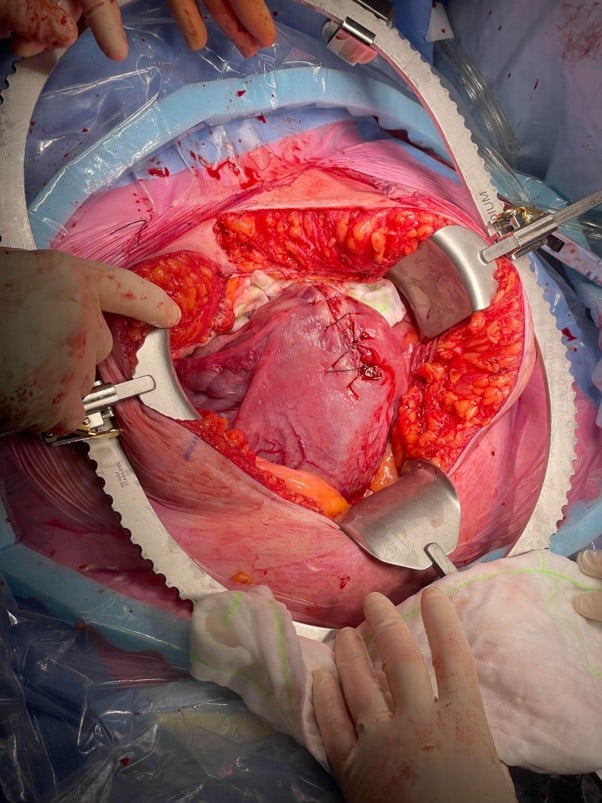
Fig. 4. Uterus after closure with locking Vicryl suture demonstrating position of vertical fundal incision superior to large surface vessels. Credit Dr Olivia Smart, Te Whatu Ora – Waitaha Canterbury.
Postoperative haemoglobin was 103 g/L from a starting point of 113 g/L. Vaginal bleeding was light postoperatively and then resolved. The patient’s recovery in hospital was straightforward, and she was discharged on day seven postpartum. Her last follow up with the obstetric service was four months postpartum. She described a full recovery to her physiological baseline, but reports ruminating at times about her sudden unplanned delivery and the loss of her uterus.
Discussion
Fundal uterine incision for PAS has been described in previous case series and retrospective review of medical records, involving both transverse and vertical incisions.3,4,5 These reports are very heterogeneous in setting and surgical technique, including conserving the uterus for future fertility. Uterine incision techniques have not been compared in head-to-head trials. These limitations of available data make it difficult to draw conclusions about optimal surgical technique at caesarean hysterectomy for PAS.
Ideally, surgical planning for PAS should take place within an experienced multidisciplinary team meeting.1 The RANZCOG clinical guideline for Placenta Accreta Spectrum (C-Obs 20) suggests that during planned caesarean hysterectomy the baby should be delivered through an incision away from the placenta.1 Avoiding the placenta minimises bleeding, allows for inspection of spontaneous placental separation, and facilitates rapid haemostatic suture to close the uterine incision.2,6 The location of the uterotomy should be individualised to the placental position for each case.6 Manual removal of the placenta should be avoided, as this may trigger severe haemorrhage.1
In this case, fundal vertical incision avoided disruption of the placenta and resulted in modest intraoperative blood loss. Preoperative bleeding was the largest source of blood loss. The uterotomy was able to be extended posteriorly under vision without limitation, until the baby could be safely delivered. The superior extent of the placenta was not reported on antenatal imaging in this case. Bedside ultrasound immediately preoperatively was an important adjunct to treatment, as it was able to detect the additional surgical complexity of the placenta covering the entire anterior uterine surface.
References
- Royal Australian and New Zealand College of Obstetricians and Gynaecologists. Placenta Accreta Spectrum (PAS) (C-Obs 20).(opens in new tab) Melbourne (AU): RANZCOG; 2023.
- American College of Obstetricians and Gynecologists; Society for Maternal-Fetal Medicine. Obstetric Care Consensus No. 7: Placenta Accreta Spectrum. Obstet Gynecol. 2018;132(6):e259-e275. doi:10.1097/AOG.0000000000002983.
- Kotsuji F, Nishijima K, Kurokawa T, Yoshinaga M, Tomiyama Y, Konishi I, et al. Transverse uterine fundal incision for placenta praevia with accreta, involving the entire anterior uterine wall: a case series. BJOG. 2013;120(9):1144-9. doi:10.1111/1471-0528.12252.
- Bakacak Z, Bakacak M, Güzin K, Yazar FM, Yaylalı A, Uzkar A. An examination by year of cases applied with caesarean hysterectomy because of placenta percreta in a tertiary centre: a retrospective cohort study. Ginekol Pol. 2021;92(4):284-288. doi:10.5603/GP.a2020.0155.
- Kayem G, Davy C, Goffinet F, Thomas C, Clément D, Cabrol D. Conservative versus extirpative management in cases of placenta accreta. Obstet Gynecol. 2004;104(3):531-536. doi:10.1097/01.AOG.0000136086.78099.0f.
- Gilner JB, Deshmukh U. Evidence-Based Perioperative Management of Placenta Accreta Spectrum Disorder. Obstet Gynecol. 2025;145(6):595-610. doi:10.1097/AOG.0000000000005920.