

The journey of laparoscopy, which is now reaching single-incision and robotic surgery, began with our quest to find ways to reduce operative morbidity.1 Since those first steps were taken, gynaecological surgery with the use of minimally invasive techniques continues to change rapidly. With computerised design and microchip-controlled safety features, the laparoscopic surgeon is dependent on the equipment and needs to understand the electromechanical function of the instruments. In this changing environment, it is vital to understand the characteristics of the commonly used surgical instruments. The basic equipment essential for any laparoendoscopic procedure includes: endoscope, camera, light source, video monitor, insufflator, trocars and surgical instruments. However, there are many variants of each available.
Disposable or reusable?
The cost effectiveness of disposable versus reusable instruments is a subject of debate. The choice of the instrument is multifactorial and depends on function, reliability and cost. So, during most laparoscopic procedures, a combination of disposable and reusable instruments is used. Frequently, disposable trocars and scissors are used, while reusable instruments can be graspers, coagulation spatula/hook and needle drivers. The commonly used laparoscopic instruments are described below.
Uterine manipulators
These allow uterine positioning and expand operating space. Several uterine manipulators are available – the HUMI® (Cooper Surgical), the RUMI® (Cooper Surgical), Spackman, Cohen, Hulka, Valtchev, Pelosi and Clearview® (Endopath). Some are reusable while others are disposable. Most come with a channel to perform chromotubation; however, some (such as Hulka tenaculum and Pelosi) lack this channel. With 210˚, Clearview has the greatest range of motion in the anterior-posterior plane. Hulka tenaculum, Spackman’s and Cohen’s have a straight shaft, hindering their range of motion and limiting their use in advanced laparoscopic procedures.2
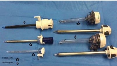
Figure 1. Trocar sleeves or collars with different textures.
Veress needle
This is a specially designed needle with a blunt-tipped, spring-loaded inner stylet and a sharp outer needle, used to achieve pneumoperitoneum while performing closed laparoscopy. It is available in disposable and reusable form, with 12cm or a 15cm length.
Most injuries in minimally invasive surgery are associated with primary port insertion, leading to an unresolved debate on the benefits of various entry techniques (open, closed or direct entry). There is no evidence that any single technique is better in preventing major vascular or visceral complications, though there is a higher risk of failed entry with closed entry. The most recent Cochrane review concluded there is a lower risk of vascular injury with the direct entry in comparison to use of Veress needle.3
Trocars/cannulas
These are used to create small passageways through the abdominal wall and are available in different textures (see Figure 1). Disposable and reusable trocars in various sizes are available and share the following common parts:
- Sharp tips cut an entry path through the abdominal wall while blunt tips stretch the tissues apart to gain access to the peritoneal cavity.
- Sleeve: is the working channel. Trocar sleeves or collars can have textures on the outer surface of the trocar that help it anchor to the abdominal wall. Some have an internal inflatable balloon at their tip and plastic/rubber ring to provide anchorage.
- Valve: different valve systems prevent gas leaking from trocars and allow the insertion of instruments.
- Side port: many trocars come with a side port that allows for gas insufflation or smoke evacuation.
Laparoscopes
The telescopes used in laparoscopy are available in sizes ranging from 2mm up to 12mm. The 10mm size is the one most commonly used in gynaecology. Similar to a hysteroscope, a laparoscope can come with an angle of view such as 0˚, 30˚ or 45˚. In an angled-view scope, the direction of vision points away from light source attachment. The 0˚ telescope offers a forward view corresponding to the natural approach and is preferred by most gynaecologists. It is useful if a less-experienced assistant is available. The 30˚ telescope can be rotated to enlarge field of view and can be advantageous for complicated cases. The 45˚ telescope is useful in single-incision laparoscopies, but is not commonly available. Every laparoscope has an engraved number by the eyepiece that specifies the viewing angle.
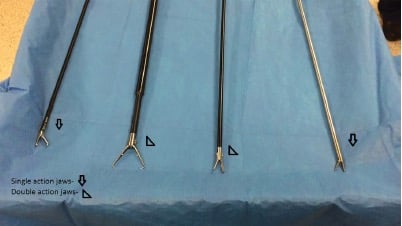
Figure 2. A range of grasper jaws.
Instrument dimensions
The commonest diameter for laparoscopic instruments is 5mm, though they range from 2–12mm. The narrower diameter (less than 5mm) instruments have less shaft rigidity and therefore are more flexible and more fragile than the wider versions. Standard instruments’ length ranges from 34–37cm. In bariatric patients or for single-site laparoscopy, 45cm-long instruments are useful.
Non-energy devices
Most laparoscopic instruments offer only four degrees of freedom of movement: in/out, up/down, left/right and rotation. In addition, certain devices called articulating/roticulating instruments offer angulation at their tips, which can be particularly useful in achieving triangulation while performing single-incision laparoscopy.4
Graspers and scissors usually have an insulated sheath, a central working device, a handle and a rotating capability at the working end.
Ringed handles are similar to the conventional ring handle found on most needle holders used in open surgery. They can be in line or directed 90˚ in relation to the working axis. Some handles are in between these two:
- a pistol handle allows integration of several functions; and
- a co-axial handle is in the instrument axis.
The handles come with different types of ratchets that provide a locking mechanism.
Scissors with curved tips, analogous to Metzenbaum, are commonly used. Most endoscopic scissors can also be attached to the electrosurgical unit. Scissors are produced with variety of tips.
Grasper jaws (see Figure 2) are either are single action (one fixed jaw and one articulated jaw) or double action (both jaws articulated). Single-action jaws close with a stronger force ideally suited for an instrument such as a needle driver. Double action allows the jaws to open wider, so they are better suited as a dissection tool. Numerous grasper variants exist, with the inner side of the jaws having different surface properties, depending on the intended use:
- Traumatic: deep serrations or toothed tip for secure grasping.
- Atraumatic: finely serrated for gentle handling.
Equally, laparoscopic tenacula are also available with single-toothed and doubletoothed jaws.
Many styles of needle drivers are available and selection largely depends on surgeon’s preference. The jaws are either curved or straight. They commonly have a flat or finely serrated grasping surface, enabling them to grasp the needle in all directions. Certain needle-holders (termed self-righting) have a dome-shaped indentation inside their jaws that automatically orientates the needle in a perpendicular direction, thus making it easier to grasp the needle. However, if there is a need to load the needle at an oblique angle, the indentation can make it harder. The needle drivers also have various types of handles (such as finger grip, palm grip, pistol grip) as described previously.
Myoma screws are in the shape of a probe with a corkscrew tip. They are frequently used during myomectomy.
The suction irrigator is a multipurpose piece of equipment. Most use a trumpet valve but some have a sliding valve. The irrigation system can be powered by various mechanisms including pressure bag or a pump. Omentum, fallopian tube or bowel can get drawn into the suction probe and care must be taken to release the attached tissues gently.
The aspiration needle is a 16/22-gauge needle used for aspiration and injection of fluids.
There are two types of knot pushers available: the closed-end and the open-end knot pusher. Both have their advantages and disadvantages.
Energy devices
Energy sources include monopolar, bipolar, advanced bipolar, harmonic, combined and morcellator devices. Monopolar devices are commonly used in endometriosis resection and for incising the vaginal cuff during laparoscopic hysterectomy. Various types of monopolar hooks and spatula are available and most scissors have an attachment to connect monopolar lead.
Bipolar devices contain the continuous waveform electrical current between the jaws of the forceps and hence reduce the chances of damage to adjacent tissue. They achieve tissue sealing and haemostasis by thermal coagulation, though they lack the ability to cut. The classic bipolar device is the Kleppinger bipolar forceps. Several types of bipolar devices, many of them in form of graspers, are now available.5
The surgical evolution of the energy devices, particularly with advanced bipolar features, has been the central point in exponential growth of laparoscopic procedures. The gain in popularity of these devices can be gauged by the fact that they are sometimes now used for open surgery and even vaginal surgery.6
Bipolar devices (such as LigaSure™, Gyrus PKS™ and EnSeal®) provide haemostasis for vessels up to 7mm. They provide a low voltage, have an impedance-based feedback that modifies the energy delivered and tissue temperature is regulated to be below 100°C. The bipolar energy thus delivered denatures the collagen and elastin in vessel walls. Denatured tissue, tissue apposition and pressure seal the vessel walls in a process called coaptive coagulation. In comparison to the traditional bipolar instruments, these devices have reduced thermal spread, diminished charring and reduced sticking. However, some of these devices require a specialist electrosurgical unit and they are costly.7
LigaSure (Covidien) provides a continuous bipolar waveform and has an integrated cutting mechanism. GyrusPK (Gyrus ACMI) delivers a pulsed bipolar waveform that allows tissue and device tip to cool during the energy off phase, but lacks the ability to cut. Enseal (Ethicon) has nanometre-sized conductive particles that direct the energy and control temperature between the jaws. Like LigaSure, it is multifunctional, with an I-Blade™ to cut the sealed tissue.
Harmonic devices have a piezoelectric crystal in their handpiece that converts the electrical energy into ultrasonic energy. This energy is delivered to the active blade at the tip of the instrument causing it to vibrate at 55 000Hz. The tip of the device cuts mechanically with a degree of collateral thermal coagulation used for haemostasis. There is no active current in the tissue. The advantage of harmonic devices is lower temperature (<80°C) as compared to other energy devices, hence reduced thermal spread and less charring. As a result of mechanical vibrations, in lower density tissue the intercellular water is vaporised at lower temperatures (<80°C) causing a ‘cavitation effect’ that can help in dissection by separating tissue layers. They are FDA approved for <5mm vessel sealing. Though harmonic devices operate at low temperatures, the active blade of the device becomes very hot and can remain so for some time. Care should be taken not to touch the vital structures with the jaws of the device for several seconds after activation.
Thunderbeat® (Olympus) combines both advanced bipolar electricity and ultrasonic energy in a single, multi-functional, handactivated instrument and can potentially reduce the surgical time.
Morcellators can be important tools for specimen removal during procedures, such as myomectomy, when a large amount of tissue is retrieved laparoscopically. Various types of morcellators are available on the market. The key safety maxim is to keep morcellator tip close to abdominal wall, to pull the tissue into the morcellator and not push the morcellator into the tissue. Morcellators require ports that are bigger than 5mm. Morcellation has recently been in news with a US Food and Drug Administration safety communication in 2014 swiftly followed by new and/or revised guidelines, including a joint statement by AGES and RANZCOG. To prevent tissue dissemination, power morcellation in an isolation bag has been proposed. Recently, an in-bag morcellation device (Alexis™ Contained Extraction System) has also been made available.8 9 10
References
- Behnia-Willison F, Foroughinia L, Sina M, McChesney P. Single incision laparoscopic surgery (SILS) in gynaecology: feasibility and operative outcomes. ANZJOG 2012; 52: 366-70.
- Van den Haak L, Alleblas C, Nieboer TE et al. Efficacy and safety of uterine manipulators in laparoscopic surgery: a review. Arch Gynecol Obstet 2015; 291.
- Ahmad G, Gent D, Henderson D et al. Laparoscopic entry techniques. Cochrane Database Syst Rev 2015; (31): CD006583.
- Berber E, Akyuz M, Aucejo F et al. Initial experience with a new articulating energy device for laparoscopic liver resection. Surg Endosc 2014; 28: 974-8.
- Harrell AG, Kercher KW, Heniford BT. Energy sources in laparoscopy. Semin Laparosc Surg 2004; 11: 201-9.
- Lakeman MM, The S, Schellart RP. Electrosurgical bipolar vessel sealing versus conventional clamping and suturing for vaginal hysterectomy: a randomised controlled trial. BJOG 2012; 119: 1473-82.
- Sankaranarayanan G, Resapu RR, Jones DB et al. Common uses and cited complications of energy in surgery. Surg Endosc 2013; 27: 3056-72.
- UPDATED Laparoscopic Uterine Power Morcellation in Hysterectomy and Myomectomy: FDA Safety Communication. Nov 2014. www.fda.gov/MedicalDevices/Safety/AlertsandNotices/ucm424443.htm .
- The Royal Australian and New Zealand College of Obstetricians and Gynaecologists. Tissue Extraction at Minimally Invasive Procedures (C – Gyn 33) Review Nov 2014.
- Cohen SL, Einarsson JI, Wang KC et al. Contained power morcellation within an insufflated isolation bag. Obstet Gynecol 2014; 124: 491-7.
One Comment
Leave a Reply


This review on laparoscopic surgical tools is incredibly informative! I appreciate the detailed descriptions and comparisons of different instruments. It’s fascinating to see how advancements in technology continue to improve surgical outcomes. Looking forward to more insights like this!