


A 39-year-old woman was referred with the problem of cyclic right inguinal pain. She had two children, aged seven and five years. The first had been delivered by caesarean section for obstructed labour, the second by elective caesarean delivery. She and her partner had been trying unsuccessfully for a third child for almost a year, with no success. The menstrual periods had become heavier and more painful over that time.
The right groin lump was generally uncomfortable, but the pain reached a crescendo just before and during menstruation. On examination, a tender inguinal swelling was palpable on the right and was initially thought to be an inflamed lymph node. The patient was otherwise healthy and well, with an unremarkable past history. Imaging suggested a hypodense discrete bilobed lesion within the inguinal canal (see Figure 1). A plan was made for laparoscopy and evaluation of fallopian tube patency then an inguinal exploration.
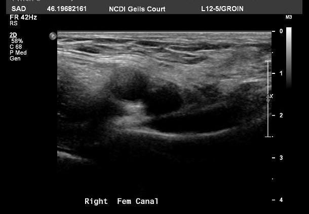
Figure 1. Imaging suggested a hypodense discrete bilobed lesion within the inguinal canal.
Laparoscopy revealed endometriosis with a large deposit of endometriosis associated with a right tubo-ovarian mass, which was excised. Within the right inguinal canal was a firm discrete mass involved in considerable scarring and adherent to the inferior epigastric vessels (see Figure 2).
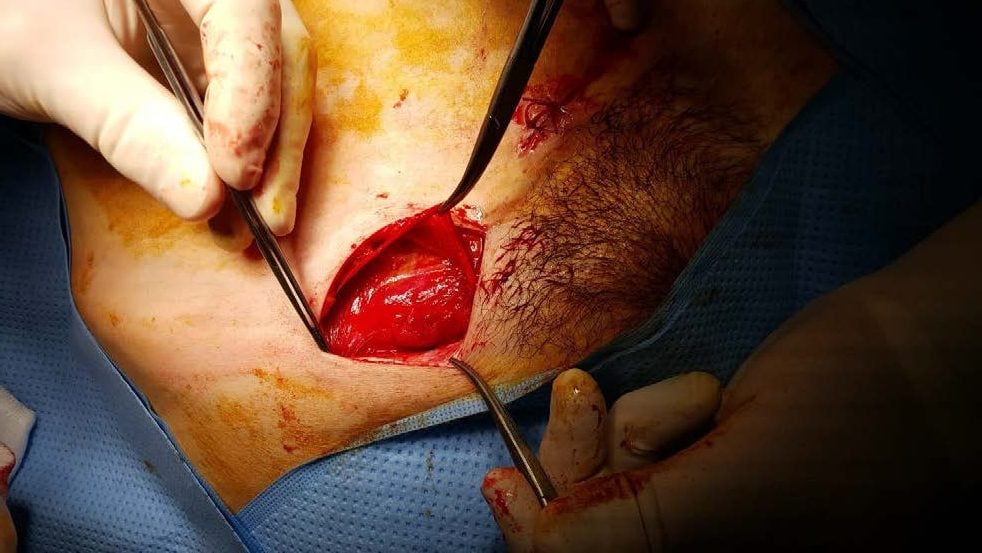
Figure 2. Dissection of the right inguinal canal following laparoscopy, revealing a dense lesion adherent to the inguinal vessels.
The lesion was carefully dissected free and he incision closed. The patient developed a groin haematoma post-operatively that was drained then settled with conservative measures. Histology revealed the inguinal lesion to be typical endometriosis.
Discussion
While pelvic endometriosis is very common, presentation of endometrial deposits outside the peritoneal cavity is very rare and can cause considerable diagnostic uncertainty. Reports have been published describing endometriosis in the diaphragm, lung and pleura, as well as the kidney and ureter, and even the extremities. Possible mechanisms for development of ectopic endometrial tissue at these sites include metaplasia and lympho-haematogenous spread.
Endometriotic deposits in the inguinal region are very uncommon, accounting for less than 0.5 per cent of all cases.1 More than 50 cases have been described in the literature.2 The typical presentation is similar to that in our patient, with cyclic pain and swelling in the groin, although some cases have been painless and in such cases the diagnosis is rarely suspected pre-operatively. Interestingly, the great majority of cases of inguinal endometriosis have been unilateral and occur on the right side as was the case in this patient.3 The reason for this is unclear, but may relate to a supposed clockwise circulation of peritoneal fluid and atypical lymphatic vessels that are more common on the right.4 5 The round ligament is often ‘protected’ by the sigmoid colon on the left and this may alter access for ectopic endometrial cells.
There is an association between inguinal endometriosis and groin hernias – typically inguinal hernias, which in themselves are less common in women – and this co-existence has been noted in up to 40 per cent of cases.6 Malignant change has also been reported 7 and one of us (PJ) has been involved in such a case in the past, and so excision of suspected inguinal endometriosis en bloc is typically undertaken. There is a strong association with intraperitoneal endometriosis, as seen in this case, so concurrent gynaecological involvement and laparoscopy is recommended whe[notn inguinal endometriosis is suspected.
References
- Hagiwara Y, Hatori M, Moriya T, et al.Inguinal endometriosis attaching to the round ligament. Australas Radiol. 2007; 51(1): 91-4.
- Licheri S, Pisano G, Erda E, et alEndometriosis of the round ligament: description of a clinical case and review of the literature. Hernia. 2005; 9(3): 294-7.
- Licheri S, Pisano G, Erda E, et al.Endometriosis of the round ligament: description of a clinical case and review of the literature. Hernia. 2005; 9(3): 294-7.
- Licheri S, Pisano G, Erda E, et al.Endometriosis of the round ligament: description of a clinical case and review of the literature. Hernia. 2005; 9(3): 294-7.
- Candiani GB, Vercellini P, Fedele L, et3. Candiani GB, Vercellini P, Fedele L, etal. Inguinal endometriosis: pathogenetic and clinical implications. Obstet Gynecol. 1991; 78(2): 191-4.
- Sataloff V, La Vorgna K, McFarland M. Extrapelvic endometriosis presenting as a hernia: clinical reports and review of the literature. Surgery. 1989; 105(1): 109-12.4. Sataloff V, La Vorgna K, McFarland M. Extrapelvic endometriosis presenting as a hernia: clinical reports and review of the literature. Surgery. 1989; 105(1): 109-12.
- Klein AE, Bauer T, Marks K, et al. Papillary clear cell adenocarcinoma of the groin arising from endometriosis. Clin Orthop Rel Dis. 1999; 361: 192-8.
2 Comments
Leave a Reply


Any recommendations for specialist in Ft. Lauderdale? 37 y/o female presenting with sudden pain and subsequent lump in groin; MRI read it as an inguinial hernia; GYN surgeon referred to General surgeon. GS physical exam led him to state it may be an endometrima rather then a hernia. L side. Onset was 10/2. Open Surgery currently scheduled for 10/24. Hx of endometriosis including laparoscopic surgery January 2018 and January 2015 (both surgeries a result of large ovarian cysts). 1 child, born vaginally in March 2017.
Would be great to know if a referral for a surgeon who could do surgery on this tripe of Endo in San Diego. Exhausted all of my options with doctors saying nothing can be done except to be put on birth control.