


Mrs F was a 36-year-old gravida 9 para 7 woman with a history of three normal vaginal births, four elective caesarean sections (CS) and a dilation and curettage (D&C) for first trimester missed miscarriage. Previous births had been complicated by postpartum haemorrhage and manual removal of placenta. Her BMI was 35 and she had a background of essential hypertension, but was otherwise well. The pregnancy in question was unplanned and Mrs F did not desire future fertility.
Mrs F was referred to acute gynaecology services by her GP, after a dating ultrasound scan (USS) suggested that the position of the gestational sac was close to the CS scar. The images were subsequently repeated and reviewed by several radiologists and obstetricians, and differing opinions emerged as to her diagnosis – live scar ectopic pregnancy versus viable intrauterine pregnancy with or without placenta accreta.
Owing to these differing opinions, further discussions between specialists and missed appointments on the patient’s behalf, Mrs F continued to be managed expectantly and regular follow-ups were arranged. During this time she developed microscopic haematuria and placenta percreta became a concern. She underwent a magnetic resonance imaging (MRI) scan at 12 weeks gestation that confirmed a lower uterine scar ectopic pregnancy, with thin myometrium and large serosal blood vessels, but no urinary bladder invasion (Figure 1). She was referred to local maternal-fetal medicine services at 13 weeks and three days, to consider medical treatment with intrasac potassium chloride (KCl) and methotrexate, or alternatively feticide before planning surgical management in order to limit the size and vascularity of the pregnancy. However, although technically feasible, this was thought to potentially increase the risk of acute haemorrhage, which would require emergency surgery.
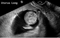
Figure 1. MRI scan at 12 weeks gestation.
A wedge resection or a hysterectomy were then proposed as treatments. As the patient did not desire future fertility and owing to her future risk for ectopic pregnancy/abnormal placentation, hysterectomy was performed at 13 weeks and four days gestation, with the legal requirements for termination of pregnancy met. The hysterectomy was performed via midline laparotomy, with cystoscopy and placement of ureteric stents at the commencement of the procedure. Intraoperative findings confirmed no bladder invasion and a highly vascular uterus of a 12–14 weeks gestation size, with an obvious defect in the low segment.
The surgery was technically challenging, and Mrs F returned to theatre twice for re-laparotomy, owing to ongoing bleeding (total estimated blood loss: 12 L), and eventually required uterine artery embolisation. She was discharged home five days postoperatively, and was followed up as an outpatient.
Clinical issues highlighted by this case include:
- The difficulty of achieving a definite diagnosis, owing to differing opinions regarding USS appearances
- A lack of evidence for optimal treatment of a second-trimester scar ectopic pregnancy.
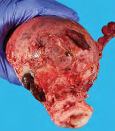
Figure 2. Hysterectomy.
Diagnostic criteria
The difficulty in diagnosing a scar ectopic pregnancy is consistently reported (with the diagnosis missed in over 13.6 per cent of cases in one review), and incorrectly diagnosed as a cervical pregnancy, spontaneous miscarriage or low intrauterine pregnancy.1 A recommended approach for reliable and reproducible criteria for diagnosing ectopic pregnancy includes all of the following2:
- Use of transvaginal USS probe at 5–12 MHz
- Visualisation of an empty uterine cavity as well as an empty endocervical canal
- Detection of the placenta and/or gestational sac embedded in the hysterotomy scar
- In early gestations (less than eight weeks) a triangular gestational sac that fills the niche of the scar – in later gestations (after eight weeks) this shape may become round or oval
- A thin (1–3mm) or absent myometrial layer between the gestational sac and the bladder
- A closed and empty endocervical canal
- The presence of embryonic/fetal pole and/or yolk sac with or without fetal cardiac activity
- The presence of a prominent vascular pattern at or in the area of a hysterotomy scar in the presence of a positive pregnancy test.
Optimal treatment
Cases of first trimester scar ectopic pregnancies are increasingly reported worldwide. Although no consensus has been reached as to the optimal treatment, several medical and surgical approaches are considered to be safe and effective, including those options that preserve fertility. There are a wide variety of primary approaches, including: intragestational methotrexate, KCI, or vasopressin; hysterosopic resection or D&C with or without adjunctive therapy, such as methotrexate; uterine artery embolisation or etoposide; and wedge resection via laparotomy.3
Cases in the second trimester are less common, and may be owing to late patient presentation or difficulty in establishing the diagnosis. As such, there is a comparative paucity of case reports to guide the choice of treatments at this gestation. Although some case series have reported conservative management with hysterotomy or D&C with or without pre-operative methotrexate or uterine artery embolisation4 5 the rates of postoperative haemorrhage were high, and subsequent emergency hysterectomy often required.6 This has led some centres to recommend expedient laparotomy and hysterectomy as first-line definitive treatment following appropriate workup and counselling.7
References
- Timor-Tritsch I, Monteagudo A. Unforeseen consequences of the increasing rate of cesarean deliveries: early placenta accreta and cesarean scar pregnancy. A review. AJOG. 2012; 207(1):14-29.
- Timor-Tritsch I, Monteagudo A, Santos R, Tsymbal T, Pineda G, Arslan A. The diagnosis, treatment, and follow-up of cesarean scar pregnancy. AJOG. 2012; 207(1): 44.e1-44.e13
- Timor-Tritsch I, Monteagudo A, Santos R, Tsymbal T, Pineda G, Arslan A. The diagnosis, treatment, and follow-up of cesarean scar pregnancy. AJOG. 2012; 207(1): 44.e1-44.e13
- Sikka P, Suri V, Chopra S, Aggarwal N. A Second Trimester Caesarean Scar Pregnancy. Case Reports in Obstetrics and Gyanecology. 2014. Article ID 828635.
- Dickerhoff L, Mahal A, Stockdale C, Hardy-Fairbanks A. Management of cesarean scar pregnancy in the second trimester: a report of three cases. J Reprod Med. 2015; 60(3-4):165-8.
- Dickerhoff L, Mahal A, Stockdale C, Hardy-Fairbanks A. Management of cesarean scar pregnancy in the second trimester: a report of three cases. J Reprod Med. 2015; 60(3-4):165-8.
- Yeo S, Tan E, Tan H. Surgical Management of a Second Trimester Scar Ectopic. Available from: eposters.rcog2015.com/e-poster/793_Yeo_Samantha_411.pdf .


Leave a Reply