

As clinicians, it is important to consider rare diagnoses when managing a busy obstetric unit. This article describes two such cases of rare uterine complications that occurred in a New Zealand tertiary obstetric unit: a uterine torsion and a uterine rupture. Uterine torsion is a rare event and is life threatening to both mother and baby if rotation is greater than 45 degrees. There are a few hundred cases reported in the literature since it was first described in 1863.1 The diagnosis can be difficult, due to vague symptoms, but prompt recognition and surgical intervention can mitigate maternal and perinatal morbidity and mortality. Uterine rupture is another rare and pathological event for both mother and fetus. This can occur prior to or during labour, and in the presence of a predisposing risk factor offers an opportunity to try to minimise risk antenatally.2 It is our hope that ongoing publications of rare cases will help improve clinician knowledge, recognition and management of rare obstetric events.
Case one: uterine torsion
A 39-year-old woman presented for an induction at 37 weeks for mild preeclampsia that was diagnosed at 36 weeks, along with a transverse lie. Her obstetric history consisted of G6P5 with all previous vaginal deliveries. On the day of induction, the lie was noted to be unstable and consent for external cephalic version (ECV) prior to induction was gained. The ECV was performed that afternoon with terbutaline tocolysis and a cooks balloon catheter was required. The woman began contracting at 0500 the next morning, the cooks catheter was removed and a small amount of dark blood noted. At 0630 the registrar was called to see the women with increasing pain. She was noted to be 1cm with a transverse lie, normal observations and normal CTG. The woman was booked for category 2 caesarean section and terbutaline was given. The woman was reviewed by registrar and consultant again at 0730 on returning from theatre with another patient and were happy to proceed with caesarean. Twenty minutes later, the registrar was called back to her room for a bradycardia. A crash caesarean section was requested, and the oncoming day registrar was instructed to perform this. General anaesthesia was given, and on opening the peritoneal cavity, a couvelaire uterus was noted with adnexal structures running diagonally over what appeared to be a twisted lower uterus. A transverse incision was made high on the posterior aspect of the lower uterine segment and the fetus was delivered via breech extraction. The placenta was delivered and the uterus was exteriorised, detorted 180 degrees and the posterior hysterotomy repaired with two-layer closure. Syntocinon bolus, syntocinon infusion and two doses of carboprost were required for tone and the total blood loss was 3100 ml. The woman recovered well postoperatively and was debriefed with her family; however, the baby was noted to have poor apgars and arterial cord gas pH of 6.76 and lactate of 17 due to the abruption. The baby required 72 hours of cooling for hypoxic ischemic encephalopathy (stage 2). During admission, the baby’s neurological exams remained normal and they had a normal on day 6 MRI brain. At the child’s 12-month clinic follow up there was normal developmental progress and growth.
The uterus can tort in both the gravid and non-gravid states and is considered pathological if beyond 45 degrees.3 The most common presenting symptom is pain with other features including malpresentation, fetal distress, vaginal bleeding and shock, and as in our case, placental abruption can be noted in 4% of cases.4 5 It can occur in acute, subacute, chronic and intermittent states. In gravid patients, if time allows, a change in placental location on ultrasound can be helpful in diagnosis.6 It can occur in all trimesters of pregnancy and postpartum with the most common being the third trimester at 49%.7 The three most recent reviews of the cases include a 1992 report (212 cases),8 2006 report (18 additional cases)9 and, most recently, a 2020 review that added another 41 cases.10 Predisposing factors include a uterine malformation, loosely suspended uterus, abdominal wall laxity, pelvic adhesions or masses including fibroids, although at times there is no pelvic abnormality identified.11
Cases associated with ECV and ongoing additional attempts at ECV with torsion-induced placental hypoperfusion and fetal distress have been described in the literature.12 Intraoperative findings include distorted lower segment, varicosities, ovaries in anterior position in relation to broad ligament and the round or infundibulopelvic ligament crossing the uterus anteriorly, and often torsion is in left to right direction.13 14 Posterior hysterotomy (intentional and unintentional) for delivery has been shown in 61% and a transverse incision shows an advantage of less blood loss, time and risk of subsequent rupture.15 For our patient, detorsion prior to hysterotomy was not possible and careful evaluation of safe entry to the uterus was performed in a timely manner. Contraception was arranged for the woman on discharge, but the subsequent rupture risk of the posterior hysterotomy is unknown and therefore advised repeat caesarean section for any future pregnancies.16 The outcome in this case was good for both mother and baby and the literature shows a decrease in both maternal and perinatal morbidity to 2.4% and 18–22% respectively since 1956.17 18 19
Case two: uterine rupture
A 35-year-old woman presented at 26 weeks to the obstetric assessment unit with sudden onset of lower back and suprapubic pain with associated vomiting. Her medical and obstetric history included a previous laparoscopic fundal myomectomy and G2P1 with a previous caesarean section three years prior for obstructed labour at 41 weeks with an incidental finding of uterine rupture at the fundus. She had been seen in the antenatal clinic with a recommendation to have an elective caesarean section at 38 weeks to avoid labour. On arrival, her blood pressure was 80/50 with a heart rate of 80. The uterus was irritable and tender to palpation, the cervix was long and closed and a bedside scan showed normal liquor volume, fetal heart rate and a breech presentation. Investigations including an urgent ultrasound were requested and the pain settled with analgesia and intravenous fluids. The ultrasound later that afternoon showed simple fluid around the right ovary and pelvis, an anterior and clear placenta and fetal weight of 940g.
The working diagnosis at this point was appendicitis versus uterine rupture and after consultant discussion, they agreed to review the patient and requested antenatal steroids to be given and a surgical review. While awaiting consultant review, a tonic contraction and bradycardia was noted by the midwife. A crash caesarean section was performed and on opening the peritoneal cavity, a 1600ml haemoperitoneum was noted with the placenta and fetus en caul in the abdominal cavity. The uterus on inspection had a defect in the fundus (see Figure 1) and this was repaired in three layers and 2 units of red cells transfused. The woman was debriefed postoperatively and had an uncomplicated recovery. The baby had a prolonged hospital course with need for ongoing follow up for chronic lung disease but was developmentally normal and was discharged from the paediatric service at two years of age.
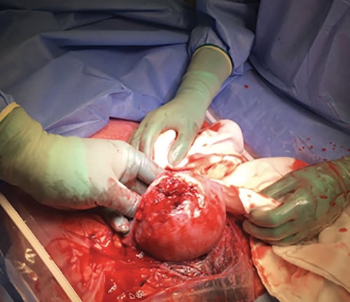
Figure 1. Fundal uterine rupture through myomectomy scar.
Uterine rupture is an obstetric emergency and often presents with an abnormal CTG, abdominal pain, loss of uterine contractions, recession of fetal parts and shock.20 Previous uterine surgery such as myomectomy or caesarean section are predisposing risk factors for uterine rupture. The incidence of rupture following myomectomy is difficult to establish in the literature, although a 2016 systematic review of 1034 pregnancies after myomectomy suggested a prelabour incidence of 1.52% and intrapartum of 0.47% which is similar to incidence for those with a prior caesarean section.21 There appeared to be no significant difference in rupture risk with fibroid location, surgical approach and technique, though the case numbers are small.22 Rupture has been shown to occur at any gestation, but most commonly in the second or early third trimester, such as our case.23 Repair of defects, obtaining haemostasis and avoidance of hysterectomy is the goal in treatment and optimal technique has not been well established due to rarity, variability in location and damage extent.24 25 The maternal mortality rate is 1/500 uterine ruptures with a perinatal mortality rate associated with rupture of 5–26%.5 Recurrent rupture data is also based on small case numbers, with a risk of 0–33%. Longitudinal ruptures show increased risk for recurrence.26 Some studies suggest that a trial of labour could be a safe alternative, preferentially to those with intramural or pedunculated myomectomy as the rate of rupture has been shown to be similar to prior caesarean; however, this is controversial and based on small case numbers.27 The American College of Obstetrics and Gynaecology recommends an elective caesarean section for those with previous myomectomy at 37–38+6 weeks and 36+0–37+0 weeks for a previous rupture.28
Both of these rare uterine events can have significant impacts for both maternal and fetal health. It is important to be wary of a patient with a previous uterine rupture, even at early gestations in subsequent pregnancies, and have a clear plan for delivery timing. Awareness of uterine torsion as a condition and recognition of intraoperative findings to allow safe hysterotomy and delivery is also valuable for obstetric clinicians. Ongoing publication of case studies aims to improve recognition of challenging diagnoses and outcomes for future patients that require swift management.
References
- Ramseyer A, Whittington J, Resendez V, et al. Torsion in the Gravid and Nongravid Uterus. Obstetrical & Gynecological Survey. 2020;75(4):243-52.
- Gambacorti-Passerini Z, Gimovsky A, Locatelli A, Berghella V. Trial of labor after myomectomy and uterine rupture: a systematic review. Acta Obstetricia et Gynecologica Scandinavica. 2016;95(7):724-34.
- Ramseyer A, Whittington J, Resendez V, et al. Torsion in the Gravid and Nongravid Uterus. Obstetrical & Gynecological Survey. 2020;75(4):243-52.
- Ramseyer A, Whittington J, Resendez V, et al. Torsion in the Gravid and Nongravid Uterus. Obstetrical & Gynecological Survey. 2020;75(4):243-52.
- Jensen JG, Uterine torsion in pregnancy. Acta Obstet Gynecol Scand. 1992;71(4):260-5.
- Ramseyer A, Whittington J, Resendez V, et al. Torsion in the Gravid and Nongravid Uterus. Obstetrical & Gynecological Survey. 2020;75(4):243-52.
- Jensen JG, Uterine torsion in pregnancy. Acta Obstet Gynecol Scand. 1992;71(4):260-5.
- Jensen JG, Uterine torsion in pregnancy. Acta Obstet Gynecol Scand. 1992;71(4):260-5.
- Wilson D, Mahalingham A, Ross S. Third Trimester Uterine Torsion: Case Report. Journal of Obstetrics and Gynaecology Canada. 2006;28(6):531-535.
- Ramseyer A, Whittington J, Resendez V, et al. Torsion in the Gravid and Nongravid Uterus. Obstetrical & Gynecological Survey. 2020;75(4):243-52.
- Ramseyer A, Whittington J, Resendez V, et al. Torsion in the Gravid and Nongravid Uterus. Obstetrical & Gynecological Survey. 2020;75(4):243-52.
- Ramseyer A, Whittington J, Resendez V, et al. Torsion in the Gravid and Nongravid Uterus. Obstetrical & Gynecological Survey. 2020;75(4):243-52.
- Ramseyer A, Whittington J, Resendez V, et al. Torsion in the Gravid and Nongravid Uterus. Obstetrical & Gynecological Survey. 2020;75(4):243-52.
- Jensen JG, Uterine torsion in pregnancy. Acta Obstet Gynecol Scand. 1992;71(4):260-5.
- Ramseyer A, Whittington J, Resendez V, et al. Torsion in the Gravid and Nongravid Uterus. Obstetrical & Gynecological Survey. 2020;75(4):243-52.
- Wilson D, Mahalingham A, Ross S. Third Trimester Uterine Torsion: Case Report. Journal of Obstetrics and Gynaecology Canada. 2006;28(6):531-535.
- Ramseyer A, Whittington J, Resendez V, et al. Torsion in the Gravid and Nongravid Uterus. Obstetrical & Gynecological Survey. 2020;75(4):243-52.
- Jensen JG, Uterine torsion in pregnancy. Acta Obstet Gynecol Scand. 1992;71(4):260-5.
- Wilson D, Mahalingham A, Ross S. Third Trimester Uterine Torsion: Case Report. Journal of Obstetrics and Gynaecology Canada. 2006;28(6):531-535.
- Gambacorti-Passerini Z, Gimovsky A, Locatelli A, Berghella V. Trial of labor after myomectomy and uterine rupture: a systematic review. Acta Obstetricia et Gynecologica Scandinavica. 2016;95(7):724-34.
- Gambacorti-Passerini Z, Gimovsky A, Locatelli A, Berghella V. Trial of labor after myomectomy and uterine rupture: a systematic review. Acta Obstetricia et Gynecologica Scandinavica. 2016;95(7):724-34.
- Gambacorti-Passerini Z, Gimovsky A, Locatelli A, Berghella V. Trial of labor after myomectomy and uterine rupture: a systematic review. Acta Obstetricia et Gynecologica Scandinavica. 2016;95(7):724-34.
- Gambacorti-Passerini Z, Gimovsky A, Locatelli A, Berghella V. Trial of labor after myomectomy and uterine rupture: a systematic review. Acta Obstetricia et Gynecologica Scandinavica. 2016;95(7):724-34.
- UpToDate. Uterine rupture: After previous cesarean delivery. 2020. Available from: www.uptodate.com/contents/uterine-rupture-after-previous-cesarean-delivery.
- Pregnancy Outcome in Patients with Previous Uterine Rupture. Obstetric Anesthesia Digest. 2007;27(4):182-183.
- Pregnancy Outcome in Patients with Previous Uterine Rupture. Obstetric Anesthesia Digest. 2007;27(4):182-183.
- Gambacorti-Passerini Z, Gimovsky A, Locatelli A, Berghella V. Trial of labor after myomectomy and uterine rupture: a systematic review. Acta Obstetricia et Gynecologica Scandinavica. 2016;95(7):724-34.
- ACOG Committee Opinion No. 764


Leave a Reply